2018年8月21日星期二

Novel Imaging Technique to Diagnose Tuberculosis Within An Hour

"/

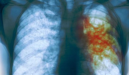
Quick diagnosis and treatment monitoring are critical due to the emergence of multidrug resistance, especially in tuberculosis. Taking this fact into consideration, a research team from Stanford University School of Medicine developed an imaging technique to diagnose tuberculosis (TB) within an hour and monitor the effectiveness of treatments. This new study has been published in Science Translational Medicine.
Dr. Jianghong Rao, the lead investigator and radiology professor from Stanford University advocated the necessity of quick TB diagnostics since the current methods take nearly two months to complete. In this duration, the disease can quickly be transmitted to other people.
This new method is easier and cheaper, compared to other current methods. Just a fluorescent microscope is enough for diagnosis along with a sample of spit from the patient.
Dr. Jianghong Rao said, “In still-developing countries where TB is most prominent, it’s hard to maintain those kinds of intensive facilities, and it can be expensive. For cases of drug-susceptible TB, the treatment success rates are at least 85%, but the rate of success is only 54% for multidrug-resistant TB, which requires longer treatments and more expensive, more toxic drugs.”
This diagnostic tool can also predict the right drug to be administered for a patient, by screening the type and number of live bacteria in the sample.
Dr Jianghong Rao is planning to get approval for this diagnostic technique from the US Food and Drug Administration (US-FDA). Dr Jianghong Rao concluded, “The hope is to make this an adaptable technology. It’s something that could be really widespread, and you wouldn’t necessarily need to use it in a hospital setting.”
from Drugdu  https://goo.gl/QgQoHk

2018年8月19日星期日

‘Orangeworm’ hacking campaign hits X-ray and MRI machines

Malware from a newly disclosed hacking campaign has infected the networks of multinational health care companies, including some X-ray and MRI machines, cybersecurity firm Symantec warned Monday.

The hacking group, dubbed Orangeworm, has hit a relatively small number of companies in more than 20 countries, Symantec said in an advisory. Nearly 40 percent of Orangeworm’s victims are in the health care industry, the advisory said. Manufacturers and IT companies that do business in health care have also been infected.
Orangeworm’s custom malware has shown up on machines that control “high-tech imaging devices such as X-ray and MRI machines,” Symantec said.
The Orangeworm revelation adds to a slew of cybersecurity challenges, including ransomware, facing the health care sector. An Indiana hospital in January paid roughly $50,000 in bitcoin to hackers that held its computer system hostage.
Congress has taken notice of the sector’s vulnerabilities. House lawmakers on Friday issued a request for information asking industry for advice on securing old hospital equipment from hacking.
Orangeworm can exploit such outdated technology by spreading across older operating systems like Windows XP, according to Symantec. “Older systems like Windows XP are much more likely to be prevalent within [the healthcare] industry,” the firm said.
Like many persistent hackers, Orangeworm has preyed on the supply chain to reach a target. “Known victims include healthcare providers, pharmaceuticals, IT solution providers for healthcare and equipment manufacturers that serve the healthcare industry, likely for the purpose of corporate espionage,” Symantec said.
Orangeworm’s malware hasn’t evolved much since its discovery and “attackers have been able to reach their intended targets despite defenders being aware of their presence within their network,” the advisory said.
Symantec referred to Orangeworm as a “group” throughout the advisory, but also said that it could be just one person. There is no indication that the hacking is affiliated with a nation-state, the firm said.
Whoever it is, they don’t seem too worried about being caught.
“Despite modifying a small part of itself while copying itself across the network as a means to evade detection, the operators have made no effort to change the [command and control] communication protocol since its first inception,” Symantec said.

from Drugdu  https://goo.gl/QgQoHk

2018年7月29日星期日

New Stroke Imaging Technology could Help Treat Brain Damage Complications Early

"/

Cone beam imaging, a new stroke imaging technology was showcased at the Society of NeuroInterventional Surgery’s (SNIS) 15th Annual Meeting in a presented study which aims at preventing nearly an hour of delay in patient care, providing them with the prospect of complete recovery.
The study, New Multiple CT Assessment of Acute Stroke Patients: Are We Ready for Prime Time?, says that new developments in imaging software in the angiosuite can give neurointerventionalists the information needed to diagnose large vessel occlusion (LVO) for an endovascular thrombectomy (EVT) in patients. The research also recommended that stroke patients could go for the angiosuite for imaging and suitable care in the future, instead of a CT scan or an emergency department.
Cone beam imaging enables precise identification of hemorrhage, occlusion site, ischemic core, and at-risk tissue. The researchers would prefer basic imaging to be done in the angiosuite using cone beam imaging. This variant of imaging gives a total 3D views of vital anatomical areas and superior resolution images compared to other methods.
Toronto Western Hospital interventional clinical research technologist and lead author of the study, Nicole Cancelliere said: “By using this technology in the angiosuite, hospitals can reduce intra-facility transfer delays and hence the time of stroke symptom onset to treatment, which will significantly reduce brain damage and improve outcomes for patients.
from Drugdu  https://goo.gl/QgQoHk

2018年7月19日星期四

New 3D Color X-Rays to Diagnose Cancer, Heart Disease and More

"/

The first-ever 3D, color X-rays were tested on a human by New Zealand Scientists, using technology that promises to enhance medical diagnostics in oncology, cardiology, neurology and orthopedics.
The scanner incorporates the Medipix3RX detector chip, a particle-tracking technology built for the CERN Large Hadron Collider, based on the traditional black-and-white X-ray technology, which was a product of the Medipix3 Collaboration, comprising CERN in Geneva and 18 other research institutions worldwide.
Phil Butler, a physics professor at the University of Canterbury in Christchurch, N.Z. said, The machine’s small pixels and accurate energy resolution meant that this new imaging tool is able to get images that no other imaging tool can achieve,”
Researchers were able to detect varied cell lines in cancerous tissue in preclinical trials on animals at universities in New Zealand, Europe and the United States, Anthony Butler said in a phone interview. The scanner could also measure arterial plaque non-invasively thereby identifying the risk of heart attack or stroke in patients and track their progress post-treatment, he added. 
Bone diseases such as arthritis and osteoporosis can also be studied using this scanner. In the coming months, a clinical trial of orthopedic and rheumatology patients is planned in New Zealand. The CERN says that the images not only very clearly differentiate between bone, muscle and cartilage, but also show the position and size of cancerous tumors.
“This color X-ray imaging technique could produce clearer and more accurate pictures and help doctors give their patients more accurate diagnoses,” said a CERN statement.

from Drugdu  https://goo.gl/QgQoHk