2018年7月29日星期日

New Stroke Imaging Technology could Help Treat Brain Damage Complications Early

"/

Cone beam imaging, a new stroke imaging technology was showcased at the Society of NeuroInterventional Surgery’s (SNIS) 15th Annual Meeting in a presented study which aims at preventing nearly an hour of delay in patient care, providing them with the prospect of complete recovery.
The study, New Multiple CT Assessment of Acute Stroke Patients: Are We Ready for Prime Time?, says that new developments in imaging software in the angiosuite can give neurointerventionalists the information needed to diagnose large vessel occlusion (LVO) for an endovascular thrombectomy (EVT) in patients. The research also recommended that stroke patients could go for the angiosuite for imaging and suitable care in the future, instead of a CT scan or an emergency department.
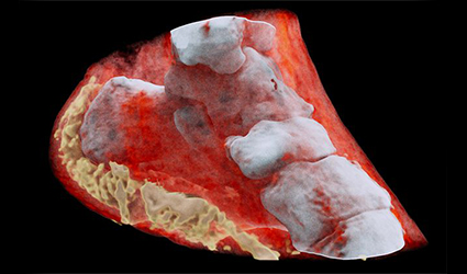
Cone beam imaging enables precise identification of hemorrhage, occlusion site, ischemic core, and at-risk tissue. The researchers would prefer basic imaging to be done in the angiosuite using cone beam imaging. This variant of imaging gives a total 3D views of vital anatomical areas and superior resolution images compared to other methods.
Toronto Western Hospital interventional clinical research technologist and lead author of the study, Nicole Cancelliere said: “By using this technology in the angiosuite, hospitals can reduce intra-facility transfer delays and hence the time of stroke symptom onset to treatment, which will significantly reduce brain damage and improve outcomes for patients.
from Drugdu  https://goo.gl/QgQoHk

2018年7月19日星期四

New 3D Color X-Rays to Diagnose Cancer, Heart Disease and More

"/

The first-ever 3D, color X-rays were tested on a human by New Zealand Scientists, using technology that promises to enhance medical diagnostics in oncology, cardiology, neurology and orthopedics.
The scanner incorporates the Medipix3RX detector chip, a particle-tracking technology built for the CERN Large Hadron Collider, based on the traditional black-and-white X-ray technology, which was a product of the Medipix3 Collaboration, comprising CERN in Geneva and 18 other research institutions worldwide.
Phil Butler, a physics professor at the University of Canterbury in Christchurch, N.Z. said, The machine’s small pixels and accurate energy resolution meant that this new imaging tool is able to get images that no other imaging tool can achieve,”
Researchers were able to detect varied cell lines in cancerous tissue in preclinical trials on animals at universities in New Zealand, Europe and the United States, Anthony Butler said in a phone interview. The scanner could also measure arterial plaque non-invasively thereby identifying the risk of heart attack or stroke in patients and track their progress post-treatment, he added. 
Bone diseases such as arthritis and osteoporosis can also be studied using this scanner. In the coming months, a clinical trial of orthopedic and rheumatology patients is planned in New Zealand. The CERN says that the images not only very clearly differentiate between bone, muscle and cartilage, but also show the position and size of cancerous tumors.
“This color X-ray imaging technique could produce clearer and more accurate pictures and help doctors give their patients more accurate diagnoses,” said a CERN statement.

from Drugdu  https://goo.gl/QgQoHk